
Can Migraine Accelerate Brain Aging According to Emerging Research
Research Shows Migraines May Be Linked To Faster Brain Aging
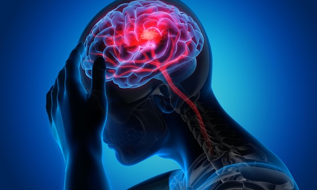
Recent findings suggest that migraine is not merely a transient pain disorder but may also signal accelerated brain aging. Neuroimaging and longitudinal studies reveal structural and functional changes in the brain of migraine sufferers that resemble early markers of neural degeneration. These include cortical thinning, white matter disruption, and vascular irregularities. The evidence points toward neuroinflammatory and oxidative stress pathways as key drivers. For clinicians, this connection underlines the importance of monitoring cognitive and vascular health in chronic migraine patients to mitigate long-term neurological risks.
Understanding the Relationship Between Migraine and Brain Aging?
The relationship between migraine and brain aging involves complex biological interactions that extend beyond headache episodes. Repeated attacks may gradually alter neuronal networks, leading to measurable structural consequences detectable through advanced imaging.
Neurological Mechanisms Linking Migraine to Brain Structure Changes
Recurrent migraine attacks appear to induce microstructural alterations in cortical thickness and white matter integrity. These changes often localize to regions involved in sensory processing, such as the occipital and temporal cortices. Chronic neuroinflammation has been proposed as a mediator of these effects, with elevated cytokine levels promoting oxidative stress that accelerates neuronal wear. Functional MRI studies demonstrate disrupted connectivity within pain-processing networks like the default mode and salience networks, suggesting long-term reorganization of neural circuits that parallels patterns seen in aging brains.
The Role of Vascular Dysfunction in Migraine-Related Brain Aging
Vascular dysfunction plays a central role in linking migraine to cerebral aging. Endothelial impairment reduces nitric oxide bioavailability, diminishing vascular flexibility and leading to microvascular instability. Chronic hypoperfusion observed in some migraine patients may cause subtle ischemic damage over time, particularly in subcortical regions rich in small vessels. Researchers have proposed that vascular biomarkers—such as circulating endothelial microparticles or altered flow-mediated dilation—could serve as early indicators of accelerated cerebral aging among individuals with frequent migraines.
Emerging Evidence from Neuroimaging and Longitudinal Studies
Growing access to high-resolution imaging has allowed scientists to trace how migraines reshape the brain across time. These observations provide compelling evidence for both structural and cognitive parallels between chronic migraineurs and older adults experiencing age-related decline.
Findings from MRI-Based Research on Migraine Patients
MRI-based research consistently reports increased prevalence of white matter hyperintensities (WMHs) among people with migraines compared with controls. These lesions often appear in periventricular or deep white matter zones associated with small vessel disease. Notably, differences between migraine with aura (MwA) and without aura (MwoA) suggest distinct neurobiological mechanisms—MwA tends to show greater occipital involvement linked to cortical spreading depression phenomena. Correlations between lesion load and mild cognitive decline further reinforce the notion that repeated migraine activity may mimic features of premature brain aging.
Longitudinal Data on Cognitive Function and Structural Brain Changes
Long-term follow-ups reveal subtle but measurable declines in executive function among chronic migraine sufferers, particularly those with high attack frequency over several years. Progressive gray matter loss has been documented in regions such as the insula, anterior cingulate cortex, and prefrontal cortex—areas integral to pain modulation and decision-making. The cumulative burden of recurrent attacks seems proportional to both structural deterioration rate and cognitive performance drop, implying dose-dependent neurobiological consequences.
Biological Pathways Potentially Driving Accelerated Brain Aging in Migraineurs
The biological underpinnings behind this phenomenon involve overlapping inflammatory, oxidative, genetic, and epigenetic pathways that compromise neuronal resilience over time.
Inflammatory and Oxidative Stress Mechanisms
Elevated pro-inflammatory cytokines like IL‑6 and TNF‑α heighten neuronal vulnerability by disrupting synaptic stability. Oxidative stress contributes additional strain by impairing mitochondrial energy metabolism and reducing cellular repair efficiency. Chronic activation of glial cells sustains this cycle of inflammation, promoting low-grade neurodegeneration similar to what occurs during normal aging but at an accelerated pace among frequent migraineurs.
Genetic and Epigenetic Factors Modulating Susceptibility
Certain gene polymorphisms involved in vascular tone regulation (e.g., MTHFR or NOS3 variants) may predispose individuals both to migraines and faster neural decline due to impaired endothelial function or heightened oxidative load. Epigenetic modifications triggered by chronic stress can further alter expression of neuroprotective genes over time, influencing how neurons respond to recurring nociceptive stimuli. Studying these gene–environment interactions could help explain why some patients experience marked cognitive effects while others remain relatively unaffected despite similar clinical histories.
Clinical Implications for Neurology Practice and Research Directions
For neurologists, linking migraines with brain aging reframes patient management from symptom control toward long-term neuroprotection strategies.
Assessing Brain Health in Patients with Chronic Migraine
Integrating advanced MRI techniques such as diffusion tensor imaging or susceptibility-weighted imaging into clinical evaluation allows early detection of subtle white matter or vascular changes before overt symptoms arise. Periodic cognitive testing targeting attention, memory, and executive domains can reveal functional decline even when imaging findings remain mild. Identifying high-risk subgroups—such as those with prolonged aura or frequent attacks—supports tailored interventions aimed at preserving brain health.
Future Research Priorities on Migraine and Neurodegeneration Linkages
Future research should prioritize large-scale longitudinal cohorts capable of establishing causality between migraine frequency patterns and objective markers of cerebral aging. Combining multimodal imaging data with biomarker profiling (inflammatory panels, oxidative markers) will refine mechanistic models describing how repeated pain episodes translate into structural decay. Another critical direction involves testing whether preventive treatments like CGRP antagonists or neuromodulation therapies can slow neural aging trajectories by reducing attack burden or mitigating inflammatory cascades.
FAQ
Q1: Can migraines permanently damage the brain?
A: While most migraines do not cause direct injury, recurrent episodes are linked with small white matter lesions that may reflect cumulative microvascular stress over time.
Q2: Are people with aura more at risk for brain aging?
A: Yes, studies show those experiencing aura exhibit greater occipital cortical changes consistent with higher susceptibility to structural alterations.
Q3: How does oxidative stress contribute to this process?
A: Oxidative molecules disrupt mitochondrial energy balance, weaken cellular repair systems, and accelerate neuron loss similar to mechanisms seen in normal aging.
Q4: Can lifestyle modifications reduce these risks?
A: Regular physical activity, balanced diet rich in antioxidants, stable sleep patterns, and stress management have shown potential benefits for both migraine control and overall brain health.
Q5: What future diagnostic tools might improve detection?
A: Emerging methods like quantitative susceptibility mapping combined with blood biomarker assays could soon allow earlier identification of migraine-related neural aging patterns before clinical symptoms manifest.